Surface modified slides let IHC laboratories do more with less
Immunohistochemistry (IHC) has come a long way since the first study using fluorescent conjugated biomarkers was published in 1942.1 What once was an experimental methodology limited to academia has now become a routine procedure in laboratories and hospitals worldwide, helping pathologists more effectively diagnose and treat patients. Innovations in IHC, such as polymer-based detection systems, automated instruments, and improved antibody engineering, have not only contributed to faster sample turnaround time, but have made IHC an essential tool for pathologists worldwide. However, despite the many innovations and technological advances made in IHC over the past 70 years, one element that is a vital part of the IHC process has undergone little change: the slide.
Worldwide, IHC continues to grow at a rapid pace and is driven by an explosion in genomic sequencing technology, global emphasis on improved healthcare, and an aging population. Yet, despite this global growth in IHC, problems still exist with regard to improper tissue fixation and varying tissue-processing procedures. Tissue fixation and processing procedures and protocols can lead to tissue wash off after heat-induced epitope retrieval (HIER), a vital step in any IHC procedure. A failed experiment due to post-HIER tissue sample loss not only leads to a loss of time, money, and effort on the part of the lab, but could lead to a longer diagnostic turnaround time or misdiagnosis, which may have an adverse effect on patient care.2,3
Tissue loss during immunohistochemical staining procedures may occur due to weak interaction of a tissue sample with glass surfaces. Histological methods for preparing tissue sections for microscopic examination commonly use glass slides with a modified surface chemistry to promote tissue adhesion, and are sold under many brand names. Such surface modifications typically include imparting a net positive electrical charge to the slide by treatment with agents such as aminosilane,4,5 polylysine,6-8 or other proprietary chemistries. However, even with such surface modifications, a proportion of tissues subjected to HIER are lifted, folded, damaged, or completely detached from slides after thermal treatment. Although partial lifting or folding of tissue sections may not significantly impact morphological assessment, the IHC results may be severely compromised, particularly for small samples such as needle biopsies.
Investigating the surface chemistry of several commercially available microscope slides (which are referred to as “hydrophobic” due to their water-repulsing characteristics) and slides which were “hydrophilic” in nature, we compared the ability of hydrophobic and hydrophilic slides in two functional areas common in IHC: tissue retention and reagent dispersal. Slides that display superior tissue adhesion ability and superior reagent dispersement could be of great benefit to laboratories seeking to minimize incidences of tissue samples loss and, as an added benefit, could reduce laboratory reagent costs—since greater reagent dispersal can lead to less reagent use.
Materials and methods
Several commercially available microscope slides were compared to slides that displayed hydrophilic characteristics. Slide reagent dispersal and drop contact angle were measured to assess hydrophobicity and hydrophilicity of slides. X-ray photoelectron spectroscopy (XPS) was also used to measure the density of amino groups attached to slide surfaces. Typically, a higher number of amino groups correlate to greater tissue adhesion.9
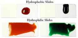
Slide hydrophilicity was measured by first hydrating slides for 10 seconds in distilled water, blotting excess water, and then depositing 200 µl of dyed tris buffered saline (TBS without detergent) onto the center of each horizontally positioned microscope slide. After five minutes, digital images of slides were captured and analyzed using ImageJ image analysis software.10 Drop outline and area of spread were recorded for each type of slide and reported as a percentage of the slide’s total working area (Figure 1).
Drop contact angle was measured by depositing 2 µl of TBS on horizontally positioned slides. Digital images of drops were captured and analyzed with ImageJ software using DropSnake plug-in according to the method of Stalder et al.10 At least 10 contact angles were measured for each slide type. Average values of contact angles for untreated, hydrophobic, and hydrophilic and corresponding standard errors were plotted.
X-ray photoelectron spectroscopy was used to measure the density of amino groups attached to slide surfaces.11,12 Samples of glass slides (2 cm x 2 cm squares) were attached to a sample holder and analyzed with a Kratos Axis Ultra XPS instrument using a monochromatic Al k-alpha x-ray source at 1486 eV and at 225 W. Spectra were acquired in the fixed analyzer transmission mode with vacuum maintained at 3.5×10⁻⁸ torr. Survey scans were taken at 80 eV pass energy between 0-800 eV. High resolution scans for N1s and C1s regions were done at 40 eV pass energy at 400 ms dwell time. Spectra were aligned relative to the C1s aliphatic carbon peak at 285 eV. Data analysis and curve fitting were performed with the Casa XPS software. Regions of interest were fitted to the synthetic component peaks with a Gaussian line shape. Relative content of amino groups was measured as an area corresponding to Gaussian peak at 399 eV.
Tissue adherence and immunohistochemistry
Fifty-six formalin-fixed paraffin-embedded (FFPE) tissues were selected that were known to be prone to detachment after HIER treatment. The ability of the different types of slides to retain tissue specimens after HIER treatment and IHC was evaluated along with tissue morphology. Each slide was mounted with serial sections. Tissue adherence was evaluated by measuring the proportion of the total tissue area that remained attached to the slide following HIER treatment. Digital images of tissues were captured and analyzed with ImageJ software to calculate the percentage of remaining tissue. Complete tissue loss was calculated as 0%, and no tissue detachment was calculated as 100% (Table 1). The HIER procedure comprised heating deparaffinized sections for 15 minutes at 121ºC in a pressure cooker with an antigen retrieval solution. All IHC reagents and antibodies were used according to the manufacturer’s instructions. Three different pathologists evaluated results independently.
| Table 1. Each tissue was assigned a percentage score reflecting the amount of tissue remaining on the slide relative to the whole tissue using Image J Software. The range of percentage scores for each type of slide was recorded. |
Results
Slide hydrophilicity was determined by analysis of drop spreading on the surface of hydrated slides. The buffer (without detergent) spread over 935.6 ±3.6 mm2 on untreated microscope slides was able to cover only 165 ±0.9 mm2 (15%) of commercially available hydrophobic slides.
The contact angles of drops formed on the surface of microscope slides were used to further assess surface characteristics,12 with smaller contact angles indicating greater hydrophilicity and larger contact angles indicating greater hydrophobicity (Figure 2). Lower contact angles encourage reagent dispersal.
| Figure 2 |
Also noted was the microscopic surface area of tissue after HIER treatment on industry-standard hydrophobic slides—specifically, the amount of tissue detachment when HIER-based methods were used to perform IHC when tissues were mounted on industry standard hydrophobic slides versus slides that were hydrophilic in nature.
Furthermore, slides that displayed a tendency to be hydrophobic experienced tissue wrinkling and damage upon retrieval by heat-based methods due to hydrophobic slides trapping water (Figure 3).
| Figure 3 |
Discussion
Stable and strong adhesion of a tissue specimen to the microscope slide surface while mitigating hydrophobic interactions is important for achieving successful and consistent sample preparation and IHC staining.
Untreated glass surfaces do not provide strong enough retention for many biological samples, and this has led to the widespread adoption of positively charged hydrophobic microscope slides for routine IHC analysis. Slides with positively charged surfaces are produced by covalent coupling of amino groups to slide surfaces and are sold commercially under a wide variety of names. Other common methods for preparing positively charged surfaces include treating slides with aminosilane or polylysine. Although these chemistries have resulted in slides with significantly improved tissue retention characteristics, there is still a need for further improvements, particularly when combined with thermal methods of antigen or nucleic acid retrieval.
Slides that display more hydrophilic properties than traditional hydrophobic slides are important in preventing tissue detachment and promoting reagent dispersal over the entire working area of the slide to ensure consistent and uniform results in IHC. A slide that can incorporate hydrophilic characteristics, prevent tissue detachment, and facilitate reagent dispersion would benefit any lab that regularly uses HIER retrieval methods in IHC or ISH applications.
- Coons AH, Creech HJ, Jones RN, Berliner E. The demonstration of pneumoccocal antigen in tissues by use of fluorescent antibody. J. Immunol. 1942;45;159-170.
- Thompson J, Scoyler R. Cooperation between surgical oncologists and pathologists. Melanocytic Pathology, 2004:36(5):496-503.
- Sompuram SR, Vani K, Messana E, Bogen SA. A molecular mechanism of formalin fixation and antigen retrieval. Am J Clin Pathol. 2004;121(2):190-199.
- Reiss M, Reiss G. Cooperation between clinicians and pathologists. Praxis (Bern 1994). 1999;88(11):471-475.
- van Prooijen‐Knegt AC, Raap AK, van der Burg MJ, Vrolijk J, van der Ploeg M. Spreading and staining of human metaphase chromosomes on aminoalkylsilane‐treated glass slides. Histochem J. 1982;14(2):333‐344.
- Metwalli E, Haines D, Becker O, Conzone S, Pantarco CG. Surface characterizations of mono‐, di‐, and tri‐aminosilane treated glass substrates. J Colloid Interface Sci. 2006;298(2): 825‐831.
- Husain OA, Millet JA, Grainger JM. Use of polylysine‐coated slides in preparation of cell samples for diagnostic cytology with special reference to urine sample. J Clin Pathol. 1980;33(3):309‐311.
- Mazia D, Schatten G, Sale W. Adhesion of cells to surfaces coated with polylysine. Applications to electron microscopy. J Cell Biol. 1975;66(1):198‐200.
- Hironori S, Masanori K, Makoto T. Adhesion of tissue sections from eleven organs on glass slides with amino group coatings. J Biosci Bioeng. 2012;113(5):661-664.
- Collins TJ. ImageJ for microscopy. Biotechniques. 2007;43(suppl 1):25‐30.
- Araujo YC, Toledo PG, Leon V, Gonzalez HY. Hydrophilicity of silane‐treated glass slides as determined from X‐ray photoelectron spectroscopy. J Colloid Interface Sci. 1995;176:485‐490.
- Stalder AF, Kulik G, Sage D, Barbieri L, Hoffmann P. A snake‐based approach to accurate determination of both contact points and contact angles. Colloids and Surfaces A: Physicochem. Eng Aspects. 2006;286:92-103.


