Biomedical research laboratories are a critical gateway between scientific discovery and real-world clinical application. As such, biomedical researchers require tools that produce high-quality data with enhanced efficiency. When it comes to imaging tools, conventional microscopes are often time-consuming, prone to user subjectivity and error, and place users at risk of ergonomic-related strain and injury. Additionally, researchers often require separate and complex software to process and analyze the captured images, which is not
practical for the demands of today’s laboratory.
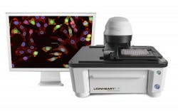
The Lionheart LX Automated Microscope (Figure 1) from BioTek Instruments integrates automaticdigital image acquisition and processing with data analysis software to simplify microscopy workflow and enable quantitative microscopy. By doing so, researchers can translate findings into the realm of practical therapeutics that can improve patient lives. The ocular-free hardware design eases ergonomic discomfort so that users can focus on the task at hand without distraction. The compact Lionheart LX offers high contrast brightfield, color brightfield, and fluorescence imaging in four channels with more than 20 available and easily changeable filter/LED cubes, along with up to 100x air and oil immersion magnification. Users can automatically image microplates, slides, cell culture dishes, and flasks.
Gen5 Microplate Reader and Imager Software controls Lionheart LX, and together they enable Augmented Microscopy for fully automated image capture, processing, and analysis. Automated features include autofocus, auto exposure, and auto-LED intensity. Automated image pre-processing optimizes images for downstream analysis, from cell counting to characterization of subcellular details. Gen5 software offers advanced image analysis including z-stacking, z-projection, montage collection, stitching, and more.
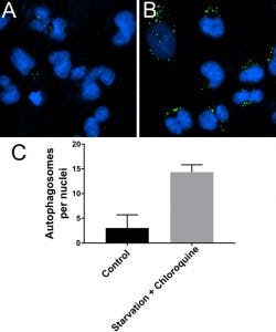
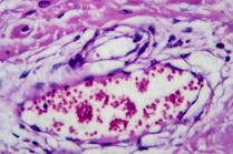
Lionheart LX is suited for a broad range of applications including histology, immunofluorescence, RNA and DNA fluorescence in-situ hybridization, and a wide range of endpoint cell imaging assays, including autophagy analysis (Figure 2). For added convenience, a quick analyze feature provides instant cell count and confluence measurements from the live camera feed; quickly finding the region of interest and displaying counts onscreen without needing to capture the image. The Lionheart LX Automated Microscope is intended for research use only.


