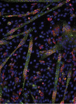
Last month’s foray into methods in molecular diagnostics discussed the basics of in-situ PCR—a potentially powerful method but one that is technically highly challenging and thus not frequently used in clinical applications. This month, we’ll look at another molecular in-situ method, but one which has significantly fewer challenges to application and is much more widely applied in clinical diagnostics. This method is fluorescent in-situ hybridization, or FISH.
Once again, we’ll start with a sample type consisting of either a slide mounted tissue section (most likely, formalin fixed paraffin embedded or “FFPE” preparation) or a slide mounted preparation of suspended cells (such as a bone marrow aspirate, or peripheral blood white cells). Ideally what we’d like for a classical FISH experiment is well separated cells with a high proportion in interphase, or on chromosomes isolated from target cells in metaphase.
(A quick refresher on interphase and metaphase, in case it’s needed: Interphase is the step in the cell cycle between cell divisions, when the chromosomal material is not actively being replicated, and is in an uncondensed form allowing for easy accessibility of chromatin and gene expression. Metaphase is the stage where chromosomes are the most physically condensed down into the characteristic paired long arms/short arms structures one associates with visual scoring and karyotyping.)
FISH in theory
The theory behind FISH is very simple, and in effect the name says it all. If we wish to observe a sample for the presence or absence of a particular nucleic acid sequence, such as a DNA region coding for a particular gene, we prepare a short single-stranded DNA probe complementary to our sequence of interest and conjugate a fluorophore of choice to the probe. We take our slide mounted sample, and if it’s intact cells or tissue, we perform a limited digestion to perforate the cell membranes enough to allow ready diffusional access of our probes into the cell nuclei for access to the chromosomal material. Then we apply our labeled probe in a small spot of hybridization buffer over the material, and after a heat denaturation step to expose the chromosomal DNA, allow the probe to selectively hybridize to its complement (if present) by incubating the reaction at the probe Tm (melting temperature, based on its length and sequence content). We follow this by a gentle washing step to remove probe material which hasn’t successfully hybridized, and place our slide under a fluorescence microscope with suitable excitation and emission filters for our probe’s fluorophore. Now all we have to do is peer in the microscope and look for the colored glowing spots which mark the presence and location of our remaining probe, and by hybridized association, the target DNA sequence of interest.
FISH in practice
If that sounds too simple to be true…that’s because it is.
If FISH is performed as described above, you’ll likely either see no signal at all when you look in the microscope (because one fluorophore label per target region isn’t bright enough for the eye to observe), or else there is a diffuse and meaningless smear of signal over all chromatin (arising from nonspecific weak hybridization of the probe to various repetitive DNA sequences).
Fortunately, neither of these problems is hard to solve. Rather than using a single probe element for our region, we can use a series of immediately sequence-adjacent probes all sharing the same fluorophore. If our probes are perhaps 20 bp in length, we can imagine selecting perhaps 20 to 40 suitable adjacent sequences in a 1 kb region of interest; we tailor the length of each probe so that its TM on associated sequence matches the rest of its cohort. By labeling these all with the same fluorophore and making them adjacent and of similar hybridization characteristics, we’ve now amplified our fluorescent signal in a small physical region enough to make it visible. (Other approaches on the signal amplification side can also be employed to resolve this; their uses will be the topic of a future article in this series).
The second problem has an even easier solution. Just as with a Southern blot, we can prehybridize our sample with short nonspecific nucleic acid “blocking agents.” These coat over and mask off the repetitive genomic sections, which have a tendency to hybridize to anything, but are readily displaced from our actual cognate target sequences by the much higher degree of sequence complementarity of the specific probes.
Let’s consider an additional bit of cleverness we can employ. Fluorophores, of course, come in limited types, particularly if you want them to work simultaneously on a single excitation filter, so you might be tempted to think this gives us a narrow palate of expressed colors to work with in our labeling here. In turn this would limit our ability to multiplex, since we wouldn’t have a way to differentiate very many different targets in one sample. The elegant solution to this is to label our short adjacent probes with different fluorophores such that they excite together but emit different wavelengths. (One could also employ a mixture of a single probe sequence but with more than one fluorophore label type). Because of their close spatial proximity, the two emitted colors combine in the observer’s eye to a new composite color. By designing probe mixtures with different ratios or mixtures of fluorophores such as 3:1, 1:1, 1:3, and so on, a highly diverse range of apparent emitted colors can be generated.
Applications of FISH
So what can we do with all of this? A first application is “chromosome painting,” where each probe mix of one apparent emitted color is specific to each chromosome. When applied to metaphase chromosome spreads, rather than the dull G-banding monochrome staining we think of, we now can light up each chromosome with a unique color. Of course, an immediate utility of this is in the detection of balanced translocations. This clinically important form of genetic rearrangement, where part of one chromosome is exchanged for another but there is no net loss or gain of genetic material, is not always easy to detect with standard karyotyping, and is effectively invisible in methods such as array CGH. With “chromosome painting” applied, however, these types of variations are immediately obvious. With full chromosome painting, this holds true regardless of whether the translocation is novel or of a commonly described variety.
Many clinical presentations, such as BCR-ABL fusion, arise from a well characterized translocation. In cases such as this, however, often a mixture of potential breakpoint donors and acceptors can be involved. The range of possible spacing between these points can make unequivocal fusion detection by classical or real-time PCR methods challenging, as multiple primer pair sets would be required to bridge all common fusion junctions. Use of the chromosome painting concept but on a smaller scale with use of just two color-coded probe regions—one for each entire donor genetic section—allows for immediate identification of a fusion between the genes regardless of the exact fusion breakpoint.
The optical appearance of a single combined color due to co-localization of probes of different colors can also be employed in FISH technique variants where the appearance of the combined color alone is enough to signal a translocation, as the immediate translocation area “lights up” in the color mixture arising from the two component regions joined. An inverse of this, known as “break-apart FISH,” can be used when a single translocation breakpoint is known to occur. By making a probe set which covers the native breakpoint but uses different probe colors on either side, the combined color is seen in normal cells, but once a breakage occurs and the two probe colors are separated, the two component colors individually appear.
FISH may also be applied to the detection of changes in copy number, where the change is large enough. For this type of application the probe set used only covers the gene or sequence of interest, not an entire chromosome. Visual (image analysis-assisted) scoring of the physical size of the probe-labeled region can be employed to determine if significant changes in hybridizing region size, either gain or loss, have occurred. Ideally this is done on mechanically extended chromosome preparations for maximum resolution, in a variation known as Fiber FISH, so named because the individual chromosomes are stretched out as distinct fibers on the slide prior to hybridization. Generation and scoring of extended DNA fibers for this approach is technically challenging, however, and array CGH is likely an easier approach where a lab is equipped to perform it for this type of analysis.
What about in-situ gene expression levels? This too can be addressed by FISH, if the probes are designed to hybridize to mRNA. If the expected mRNA copy number will be quite high, a small probe set can be employed which will generate a detectable signal from a concentration of mRNA target molecules, but will remain below apparent visibility on single-copy DNA genomic sequences. (Alternatively, sample pre-treatment with a DNase to destroy the genomic DNA sequences could be considered to ensure signal arises from RNA). In addition to mRNA, RNA FISH has been employed to examine the expression patterns of lncRNA (long, non-coding RNAs) and miRNA (short RNA molecules involved in selective post-transcriptional
regulation of gene
expression).
PCR vs. FISH
Unlike in-situ PCR, FISH is commonly used by clinical molecular laboratories. This difference is primarily due to the robustness (reliability) of FISH as opposed to in-situ PCR. As readers of last month’s column will recall, the tissue pre-treatment and permiabilization needed for in-situ PCR is a narrow balancing act between allowing access of reagents into target sequences and restricting the uncontrolled diffusion of amplicons away from site of generation. By contrast, FISH is very forgiving. The relatively large size differential between the target molecules (thousands or millions of base pairs in length) versus the 10-to-20 base pair size of the probes means that the digestion permiabilization “sweet spot” is very wide. (Note that an obvious exception to this would be the FISH for miRNAs, mentioned above. This would be expected to share the same challenges as in-situ PCR in this regard; but miRNA FISH is primarily a research tool.)
Finally, data from a successful FISH assay can be visually appealing, verging on the artistic (thus its not infrequent appearance on journal covers), and, in many applications, can be immediately understandable and interpreted by even non-specialists if a suitable figure legend is provided. The appearance of three bright colored regions when one expects two, or a markedly different color in one cell versus another, are dramatically visible to even the untrained eye.
About the Author

John Brunstein, PhD
is a member of the MLO Editorial Advisory Board. He serves as President and Chief Science Officer for British Columbia-based PathoID, Inc., which provides consulting for development and validation of molecular assays.

