Hematology automation has progressed steadily since Wallace Coulter first applied electrical impedance technology to counting red cells and white cells.1 By the 1980s, most hematology laboratories were reporting out a 7-parameter complete blood count (CBC) and three-part differential obtained from a single aspiration on a stand-alone, bench-top instrument. Eventually, this process was upgraded even further when it became possible to obtain these results without uncapping the sample. When this became state-of-the-art, CBC turnaround time (TAT) was primarily dependent on how fast a laboratorian could do a manual 100-cell differential and/or a manual reticulocyte count.
Manufacturers have developed finely tuned hematology analyzers that achieve good levels of precision and accuracy in cell counting through the examination and identification of thousands, not hundreds, of cells in each sample analyzed. The challenge of reporting precise results for immature cells using manual methods is exemplified with manual reticulocyte counts, which routinely have a CV of approximately 25%. Manual methods, even for immature cell counts, are being replaced with precise, reliable automated hematology systems that provide faster reportable results within the first aspiration.
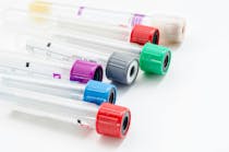
At the highest level of hematology laboratory automation are scalable, configurable automation systems dedicated to shepherding lavender (EDTA) top tubes through the following analytic determinations: CBC, 6-part white blood count (WBC) differential, nucleated red blood cell (NRBC), reticulocyte count (RET) and immature retic fraction (IRF), automated immature platelet fraction (IPF), and automated smear preparation and staining. These on-demand tests are standardized assays that meet performance goals, decrease technologist hands-on time, eliminate batch testing, and provide results faster to physicians. When this testing is supported by a hematology-specific middleware product, clinical laboratories are automatically reporting up to 85% of their test volume without any operator intervention.
Today, hematology automation platforms offer more than CBC testing from a single EDTA sample. Laboratories that have incorporated HbA1c testing on high speed hematology lines are performing > 90% of assays from lavender top tubes with minimal technologist intervention. Autovalidation of HbA1c results can run as high as 90%. Further process improvements are coming to the forefront of hematology, such as pre- and post-analytical sample sorting/archiving and automation of digital smear review. Now, these newly formed EDTA work areas can manage traditional hematology testing as well as the HbA1c traditionally tested in the chemistry department.
Automation: pre-analytical
Common processes and practices prior to analysis have also recently been reshaped. Having a single automated island for hematology testing allows sample receiving areas to quickly route samples for testing. Automation platforms can confirm sample receipt (“in-lab”) to the LIS at the point of barcode reading on the automation line, which helps reduce the need for manual intervention at the pre-analytical stage.
One area of hematology testing that was late to become automated was reagent preparation. Hematology technologists frequently have been frustrated about the time and effort required to change 20L cubes of diluent. Recent additions to automated hematology lines address this concern by including units that utilize concentrated reagent which is diluted using an in-lab water supply. This approach minimizes the time and effort required to prep the analyzers prior to the highest volume run of the day and enables laboratorians to avoid interruptions in testing due to the need to frequently change diluent in high-volume testing settings.
Automation: clinical decision making
The latest hematology technologies automatically provide results to the physician on immature cell population characteristics that can reflect the state of leukopoiesis, erythropoesis, and thrombopoiesis in the bone marrow through analysis of peripheral blood.
Reticulocyte panel. Four immature red blood cell (RBC) parameters can be automatically reported with every CBC to provide the information needed for the physician to assess the state of erythropoiesis. These parameters are the number of circulating reticulocytes (RET), IRF, hemoglobin content of reticulocytes, and nucleated red blood cells. All four of these parameters are direct measurements of erythropoiesis.
Reticulocyte hemoglobin, the latest of these parameters to become widely available, can be useful to physicians in the diagnosis of iron deficiency and iron deficiency anemia (ID/IDA) because the parameter is a direct measure that reflects the iron available for hemoglobin synthesis. In a study in 2010 by Van Wyck et al,2 this parameter was faster to obtain than chemistry tests, which demonstrated higher biological and total variability than the direct hematological measurements of reticulocyte hemoglobin. This parameter can also be of value to the physician in the diagnosis of functional iron deficiency in end stage renal dialysis (ESRD) and patients receiving r-HuEPO therapy.3-8
Immature platelet fraction. Immature platelets were first described in 1992 by Ault et al,9 who coined the term “reticulated platelets” to describe large platelets with elevated nucleic acid content. Subsequent studies have shown the analysis of IPF on automated hematology analyzers to be a stable and reproducible parameter10 compared to the measurement of reticulated platelets using flow cytometry11-13 for providing information on thrombopoiesis. IPF levels typically rise as bone marrow production of platelets increases. Therefore, their measurement provides the physician with an assessment of bone marrow thrombopoiesis from a peripheral blood sample in a way that is similar to the assessment of erythropoiesis using reticulocyte counts. IPF can be useful to physicians in the differential diagnosis of thrombocytopenia as a result of platelet destruction versus compromised production.
Automated on-demand diabetes testing. According to the American Diabetes Association, in the United States diabetes consumes $306 billion in direct medical costs, or 20% of the nation’s healthcare expenditures. This is an increase of 41% from 2007.
While there is no unique biologic marker for diabetes, the 2009 International Expert Committee recommended that Hemoglobin A1C (HbA1c) replace fasting glucose for the diagnosis of diabetes due to pre-analytic variables, analyzer bias, and diurnal variation. (For example, blood glucose levels from analyzer bias can misclassify as many as 12% of patients.14) HbA1c concentrations also correlate with the risk of developing microvasculature disease and reflect overall glycemic exposure. The International Expert Committee has determined that values of 6.0% to 6.4% identify persons at risk and indicate that treatment interventions are appropriate to delay or prevent the disease.
One way HbA1c can be measured is by using analyzers on a chemistry line. However, because a hemoglobin result is also needed, it can be more efficient to analyze the hemoglobin and HbA1c on the same automation line from the same tube, without technologist intervention. In recent years, hematology manufacturers have integrated an HbA1c testing methodology platform, HPLC, into a single EDTA automation line, enabling laboratories to conduct on-demand HbA1c testing. The adoption of this approach is increasing as more physicians follow the current testing recommendations.
Automation: standardization
Today, both small and large integrated networks and other entities having multiple hematology testing sites can achieve standardization of sample and data management. Hematology testing systems for these multi-lab operations provide the following for standardization, thereby eliminating discrepancies that may occur when a patient is tested at different laboratories: identical technology platforms; quality control procedures; calibrators and controls; and reagents.
Examination of blood films can be time-consuming and requires significant expertise. The development of cell image analysis systems that pre-classify WBC, assess RBC morphology, and estimate platelet counts addresses the need for automation in this traditionally manual testing phase. Newer hematology automation lines can integrate cell image instruments directly on the line. This not only reduces the amount of “sneaker net” required to finalize the report but aids in the standardization of cell classification.
Hematology professionals may memorize dozens of reflex or repeat testing rules for problematic samples or abnormal results. The growing use of hematology-specific middleware automates the application of these rules and reduces the number of notes that used to be found taped to hematology analyzers. Middleware is not only capable of standardizing the reflexive actions on abnormal samples, but can also be used to consolidate information from multiple labs within a healthcare network. Newer rules include look-back and even rules based on changes in sample flagging over time. Because most of the EDTA tubes can pass through the same automation line and be recorded in a middleware system, middleware can also be leveraged to provide management reports that provide information needed to optimize sample processing.
Automation: post-analytical
Sample processing after analysis also flows differently with complete automation of hematology testing. The presence of immature granulocytes in the peripheral blood typically triggers a sample for manual smear review. New hematology analyzers now report these automatically in the WBC differential as immature granulocytes, reflecting the presence of metamyelocytes, myelocytes, and promyelocytes in the sample.
This automated, reportable measurement allows samples with immature WBCs to be autovalidated using rules established by the laboratory to reduce manual smear review. The use of hematology-specific middleware still allows the hematology lab to create lab-specific rules to review samples from critical or intensive care units or chemotherapy patients based on either a change in IG concentration or a change in sample flagging.
In addition, automated hematology systems are able to archive and sort EDTA samples after testing to facilitate rule-based reflex testing or sample storage.
Automation: staff shortages
Automation in the hematology lab may play a significant role in addressing anticipated staff shortages in a number of locations. One hospital in Queens, New York, part of the New York-Presbyterian Healthcare System, performs more than 2.3 million tests per year. Its laboratory foresees significant staff shortages within the next five years. A significant number of clinical laboratory professionals are nearing retirement, and few replacements are expected.
Among 2,200 HbA1c tests per month, however, more than 95% are managed by a completely automated process. At peak workload levels only two laboratorians are needed to manage this hematology automation line, one of whom does manual CBC differentials as needed. Because HbA1c testing is integrated on the line, there is no need for a laboratorian to batch test these samples. Automation allows more than 90% of reimbursed lavender top tubes to be tested on a single line.
Automation: cross-training
“Full automation” of hematology was once envisioned in terms of a single sample-receiving station that would automatically move any sample type to the appropriate analysis station. Based on the practical experience of labs with automation, however, it now appears that hematology may not go the way of chemistry, due to the unique capabilities that will be required of hematology automation. “Full” hematology automation may be driven by the need for STAT turnaround time, the lack of centrifugation, the need for at least some review of smears by technologists, and the variety of reflex testing used in hematology. One laboratory supervisor with more than 30 years’ experience stressed recently to this writer that hematology processing does not fit the chemistry mold. She noted that the key issue is cross-training and experience with manual smear reviews. This lab leader believes that hematology specialists will still be needed to review cell morphology and release results on samples with abnormal or immature cells. [Editor’s note: MLO would be curious to know if readers who work in hematology agree or disagree: let us know what you think at mlo-online.com]
So in summary…
This review of hematology automation has described both analytic and supporting software capabilities. From the point of view of analysis, the ability to analyze and report immature forms of three cell lines (WBC, RBC, and PLT) enables the hematology laboratory to fully automate parameters that can aid physicians in their therapeutic decision-making processes. Providing information about erythropoiesis and thrombocytopenias can help the physician choose a clinical pathway and monitor therapy. New software allows for autovalidation of hematology results and immediate reporting to the LIS. Integrating the testing of HbA1c, which is growing in importance and frequency, on a hematology line instead of routing the sample to chemistry supports laboratory efficiency. “Full” automation of hematology may not follow the chemistry model—but the adoption of automation by the hematology department carries with it important implications for the management of the clinical medical laboratory as a whole.
References
- Lemelson-MIT Program. Wallace A. Coulter (1913-1998): automated blood analysis. Inventor of the Week archive. http://web.mit.edu/invent/iow/coulter.html. Accessed November 27, 2013.
- Van Wyck DB, Alcorn H Jr, Gupta R. Analytical and biological variation in measures of anemia and iron status in patients treated with maintenance hemodialysis. Am J Kidney Dis. 2010;56(3):540-546.
- Brugnara C. Schiller B, Moran J. Reticulocyte hemoglobin equivalent (Ret He) and assessment of iron-deficient states. Clin Lab Haem. 2006;28(5):303–308.
- Brugnara C, Colella GM, Cremins J, et al. Effects of subcutaneous recombinant-human erythropoietin in normal subjects – development of decreased reticulocyte hemoglobin content and iron-deficient erythropoiesis. J Lab Clin Med.1994;123(5):660-667.
- Brugnara C, Zurakowski, Dicanzio J, Boyd T, Platt O. Reticulocyte hemoglobin content to diagnose iron deficiency in children. JAMA. 1999;281(23):2225-2230.
- Brugnara C. Iron deficiency and erythropoiesis: new diagnostic approaches. Clin Chem. 2003;49(10):1573-1578.
- Saigo K, Sakota Y, Masuda Y, et al. Automatic detection of immature platelets for decision making regarding platelet transfusion indications for pediatric patients. Transfus Apher Sci. 2008;32(2):127-132.
- Ullrich C, Wu A, Armsby C, et al. Screening healthy infants for iron deficiency using reticulocyte hemoglobin content. JAMA. 2005;294(8):924-930.
- Ault KA, Rinder HM, Mitchell J, Carmody MB, Vary CP, Hillman RS. The significance of platelets with increased RNA content (reticulated platelets). A measure of the rate of thrombopoiesis. Am J Clin Pathol. 1992;98(6):637-646.
- Jung H, Jeon HK, Kim HJ, Kim SH. Immature platelet fraction: establishment of a reference interval and diagnostic measure for thrombocytopenia. Korean J Lab Med 2010;30(5):451-459.
- Kuang M, Luo Y, Yan S, Chi P. Comparison of methodologies for detecting reticulated platelets and establishment of normal reference range. Sysmex Journ Intl. 2011;20(1):1-4.
- Saigo K, Sakota Y, Masuda Y, et al. Automatic detection of immature platelets for decision making regarding platelet transfusion indications for pediatric patients. Transfus Apher Sci. 2008;38(2):127-132.
- Cremer M, Paetzold J, Schmalisch G, et al. Immature platelet fraction as novel laboratory parameter predicting the course of neonatal thrombocytopenia. Brit J Haematol. 2009;144(4) 619–621.
- Sacks D. The diagnosis of diabetes is changing: how implementation of hemoglobin A1c will impact clinical laboratories.2009. Clin Chem 55(9):1612-1614.


