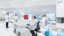
Clinical microbiology laboratories are undergoing a rapid transformation, with many of these changes posing daunting challenges. The current vacancy rate in U.S. microbiology laboratories is over 10 percent with an additional 17.4 percent of the laboratory staff projected to retire in the next five years.1 At a time when the complexity of diagnostic testing and associated costs are increasing, labs are experiencing reimbursement pressure from both government and commercial payers.11 These forces are driving laboratories to consolidate and move specimens to centralized facilities with the goal to improve testing efficiencies and decrease costs and turnaround time.
Laboratory consolidation also brings challenges. Testing is potentially moved away from physicians at a time when the interpretation of sophisticated tests requires active collaboration with the microbiology staff. Also, concentration of diagnostic procedures in large centralized laboratories poses increased safety risks for the technical staff at a time when infections with antimicrobial resistant organisms and other communicable pathogens are increasing. We can accept these changes as inevitable problems, or we can view them from another perspective. We can say necessity is the mother of innovation, or to rephrase—when they give you lemons, make lemonade. Today our innovation—our lemonade—is comprehensive automation from specimen receipt to transmission of the final report.
Automation in microbiology is not simply automating the traditional practices of processing specimens and incubating cultures—that would not be transformative. Automation should be viewed as an all-encompassing approach to processing samples and communicating information that enable laboratory technologists to perform skilled tasks in ways not historically possible. This is why automation was recently described as “a paradigm shift in clinical microbiology, representing the beginning of the future.”2
Automation in microbiology enables laboratories to do much more than eliminate the manual repetitive practices and inefficiencies of the past—the laboratory noise. Realizing the full value of automation, however, means fundamentally reexamining the entire laboratory workflow.
Specimen processing
Improved efficiency and specimen traceability are achieved with automation. Specimens are processed without delay upon receipt in the laboratory, with the automated selection of appropriate media for individual specimens, labeling the plates with barcodes, and inoculation of the media before they are transported to incubators on track systems. The accuracy of specimen inoculation is facilitated with the use of calibrated pipettes and streaking of multiple culture plates can be done simultaneously in a variety of patterns. For maximum benefits, workflow changes are required, including elimination of the practice of batching specimens, manual creation of worklists, and aligning work schedules with traditional daytime service hours. Additionally, automation challenges us to think differently about the traditional processes. We streaked plates historically in a quadrant pattern to obtain isolated colonies and perform semi-quantitation. However, this is unnecessary because automation provides more uniform, reproducible plate streaking with more isolated colonies and accurate quantitation.3-5
Automation also provides a level of safety never imagined. Techs do not have to handle most specimens with automated processing. Furthermore, since the steps of transferring culture plates to incubators, examination of cultures with digital imaging, and the automation of identification and antimicrobial susceptibility testing, it is conceivable that lab technologists may never have to handle a culture plate.
Incubation and imaging of culture plates
Full laboratory automation moves plates from the processing area to incubators, eliminating delays in incubation. The plates are incubated under ideal growth conditions of stable temperature and atmosphere because the doors are not opened, and growth is monitored through images taken by sophisticated camera systems at predetermined time periods. Thus, significant growth can be detected earlier, with improved recovery of both common and slow-growing pathogens.6-10
Imaging algorithms are capable of creating idealized images that are more than simple photographs under different lighting conditions. These images are similar to what a photographer can create with advanced photo processing software. Software also allows these images to be examined under increased magnification so subtle differences in colony morphologies or the presence of mixed cultures can be recognized. However, interpreting these images is a new skill for technologists that must be mastered. To continue physically examining plates at a workbench that includes the “art” of the sights and smells of traditional microbiology is to deny the benefits of the “science” that comes from automation.
Automation can provide a library of images for training purposes, or the images of an individual patient’s culture for discussion with a healthcare provider. Digital imaging allows a technologist to examine culture plates in a specialized reading room, at home, or remotely hundreds or thousands of miles away from where the culture is performed, enabling technologists to consult with peers or specialists as needed. Sophisticated imaging software can also determine if growth is present on a culture plate as well as the quantity of growth, thus permitting the technologist to concentrate on processing plates with significant microbial growth.
Workup of culture plates
Again, to realize the value the imaging, digital imaging times must be selected to optimize detection of significant growth as early as possible and, more importantly, the processing of the cultures for identification (ID) and antimicrobial susceptibility testing (AST) must be initiated at the time the imaging is performed. There is little value in imaging the culture plates and then delivering the plates to an external holding area or “output stack” where the plates remain for hours before further processing.
The full benefits of automation require additional changes in traditional practices. The day starts for most traditional labs by distributing all incubated culture plates to specific desks (e.g., urine, stool, respiratory, wound, etc.). This offers the advantage of performing repetitive work in a standardized manner and facilitates teaching the processing of specific specimen types. But this also fosters inefficiencies such as uneven distribution of work across workbenches, a lack of checks and balances on the quality of work performed, and the difficulties in observing the complete diagnostic picture for an individual patient.
With automation, each culture can be incubated at a predetermined time from the initial processing, so culture plates are ready for further processing throughout the day and evening. This processing is most efficient—and informative—when an individual desk processes all specimen types, allowing a technologist to see multiple specimen types from an individual patient.
The technologists who examine images from culture plates determine which colonies represent significant growth. This skilled task may not be replaced by automation. However, the actual picking of selected colonies, preparation of a standardized inoculum, and inoculation of MALDI plates for ID testing and AST panels can be automated. The results of the ID and AST test must also be interpreted by skilled technologists, so it is important that the technologists work as a coordinated team. There is also the opportunity with today’s automation and the computerized analysis of lab data, to supplement interpretation of patient data with trend analysis of infection patterns and antimicrobial resistance.
Automation may also improve laboratory operations by providing metrics to measure the efficiency of the laboratory and individual technologists. Automated systems can track the timing of all specimens as they move through the diagnostic pathway and determine when work will need to be performed and when results can be reported.
The economics of total lab automation
Finally, as we look at total lab automation, the question has frequently been asked is, “Can we afford it?” Clearly automation is a significant investment, but the more appropriate question is posed by Thomson and McElvania,2 “Can you afford not to automate?”
In an era when laboratories have to reduce costs, improve efficiency and quality, and provide more timely, accurate results to inform patient management, automation is the only solution. Thomson and McElvania2 demonstrated they were able to reduce staffing for both processing specimens and working up cultures, decrease time to results and costs by performing fewer subcultures and earlier reading of cultures, and increase the number of specimens processed by each technologist by using automation to eliminate inefficiencies. In their model, they were able to demonstrate a return on investment in three years rather than their projected five-year ROI through labor savings alone.
Yes, there is an initial investment in automation and the infrastructure to support automation. Yes, successful implementation of automation requires changing traditional workflow processes. Yes, strong leadership and teamwork will be needed through the transition. But the rewards are great: Improved efficiencies, more timely results for better patient management, and more cost-effective diagnostics. Automation truly is the future of clinical microbiology.
REFERENCES
- Garcia E, Kundu I, Kelly M, Soles R. 2019. The American Society for Clinical Pathology’s 2018 vacancy survey of medical laboratories in the United States. Am J Clin Pathol 152:155-168.
- Thomson R, McElvania E. 2019. Total laboratory automation: what is gained, what is lost, and who can afford it? Clin Lab Med 39:371-389.
- Croxatto A, Dijkstra K, Prod’hom G, Greub G. 2015. Comparison of the InoqulA and WASP automated systems with manual inoculation. J Clin Microbiol 53:2293-2307.
- Iversen J, Stendal G, Gerdes C, Meyer C, Ostergaard C, Frimodt-Moller N. 2016. Comparative evaluation of inoculation of urine samples with the Copen WASP and BD Kiestra InoqulA instruments. J Clin Microbiol 54:323-332.
- Croxatto A, Marcelpoil R, Orny C, Morel D, Prod’hom G, Greub G. 2017. Towards automated detection, semi-quantification, and identification of microbial growth in clinical bacteriology: a proof of concept. Biomed J 40:317-328.
- Lainhart W, Burnham C. Enhanced recovery of fastidious organisms from urine culture in the setting of total laboratory automation. J Clin Microbiol 56(8):e00546-18.
- Klein S, Nurjadi D, Horner S, Heeg K, Zimmermann S, Burckhardt I. 2018. Significant increase in cultivation of Gardnerella vaginalis, Alloscardovia omnicolens, Actinotignum schaalii, and Actinomyces spp in urine samples with total laboratory automation. Eur J Clin Microbiol Infect Dis 37:1305-1311.
- De Socia G, Di Donato F, Gabrielli C, Belati A, Rizza G, Savoia M, Repetto A, Cenci E, Mencacci A. 2018. Laboratory automation reduces time to report of positive blood cultures and improves management of patients with bloodstream infection. Eur J Clin Microbiol Infect Dis 37:2313-2322.
- Burckhardt I, Last K, Zimmermann S. 2019. Shorter incubation times for detecting multidrug resistant bacteria in patient samples: defining early imaging time points using growth kinetics and total laboratory automation. Ann Lab Med 39:43-49.
- Bailey A, Burnham C. 2019. Reducing the time between inoculation and first-read of urine cultures using total lab automation significantly reduces turn-around time of positive culture results with minimal loss of first-read sensitivity. Eur J Clin Microbiol Infect Dis 38:1135-1141.
- Stone J. 2018. With Reduced Reimbursement from Medicare, Anatomic Pathology Groups and Clinical Laboratories Must Learn to Optimize Collections from Managed Care Payers to Stabilize Financials and Survive the Industry Shift. Dark Daily.


